A new trend is a word used not just for technology and fashion but in the field of medicine as well. The discovery and progression of medical tools really brought us comfort and relief. One of which is the use of high energy radiation or medical imaging in the diagnosis and treatment of diseases. This is known to as Radiology. If you want to know more about this procedure, visit imagingassociates website.
Variety of Imaging Techniques
The separation of radiology and radiation oncology give way to the birth of this two distinct specialties. Most people don’t really know the difference between diagnostic imaging and interventional radiology.
To further discuss this, diagnostic imaging is the use of noninvasive methods for knowing and monitoring diseases and injuries. This is through the production of images that show the internal structures and organs of the patient’s body. While interventional radiology or IR is a medical practice that gives less invasive image-guided diagnosis and treatment of diseases.
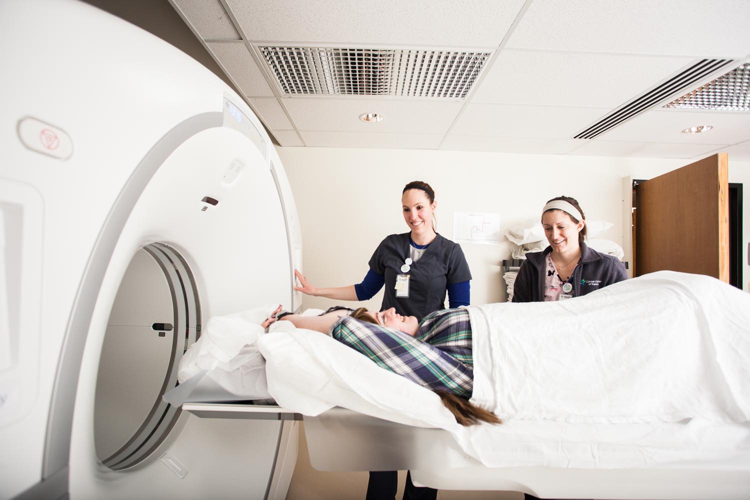
Diagnostic imaging techniques include X-rays, fluoroscopy, CT scans, MRI and radiofrequency electromagnetic radiation to detect and diagnose diseases anywhere in the body. These procedures cover plain film examinations, gastrointestinal studies, genitourinary studies, arthrograms, hysterograms and others. It’s called noninvasive because the patient’s body is not cut open and entered by any kind of equipment for imaging. Though some procedures integrate diagnostic imaging to minimal invasive procedures to diagnose and treat diseases
IR, on the other hand, is a procedure done using small instruments that enter the body through a tiny skin nick or incision. Since it’s minimally invasive, it has been labeled as the surgery of the millennium. Treatment of benign tumors, certain types of cancer, uterine fibroids, spine fractures, and pelvic pain can now be possible through IR. Some of the techniques done in IR include:
- Angiography
- Arteriovenous Malformations (AVM)
- Balloon Angioplasty
- Central Venous Access
- Chemoembolization
- Embolization
- Gastrostomy Tube
- High Blood Pressure
- Needly Biopsy
- Radiofrequency Ablation
- Stent
- Urinary Tract Obstruction
- Uterine Artery Embolization (UAE)
- Uterine Fibroid Embolization (UFE)
- Varicocele Embolization
- Vena Cava Filter
- Vertebral Augmentation
The Patients Preparation and Readiness
To produce a good quality image, the patients may be required a specific preparation depending on the procedure. The doctors may need the patients to do as follows:
- Fasting – Patients mustn’t eat or drink anything for a certain number of hours before the procedure.
- Drink a certain amount of fluid (Depends on the procedure the patient must take it)
- Wear comfortable clothes – The patient must not wear anything with zippers, metallic buttons, and snaps and even jewelry (something that may interfere with the scan).
To avoid any danger to the patient, the doctors must ask the patient if he/she is taking some medication or have some allergy to certain drugs.
If you want to meet experts and friendly radiologist, see http://imagingassociates.net.au/.